

You know that moment when your dentist casually mentions, “Let’s take a quick X-ray”? Maybe you’ve wondered what exactly they’re looking for, or if all that imaging is really necessary. We get it. Dental X-rays can feel like one of those mysterious parts of dental care that nobody really explains.
Here’s the thing: these images are doing so much more than you might realize. While we’re examining your teeth during a routine visit, there’s a whole world beneath the surface that our eyes just can’t see. Cavities hiding between teeth, infections brewing at the roots, bone loss happening silently – dental X-rays reveal what’s actually going on in there before small problems turn into painful (and expensive) ones.
We’ve seen firsthand how these diagnostic tools change everything. They help us catch issues early, plan treatments with precision, and keep your oral health on track. And with modern technology, they’re safer and faster than ever before. Let’s walk through what dental X-rays really are, why we rely on them, and what you can expect when it’s time for your next image.
What Are Dental X-Rays and How Do They Work?
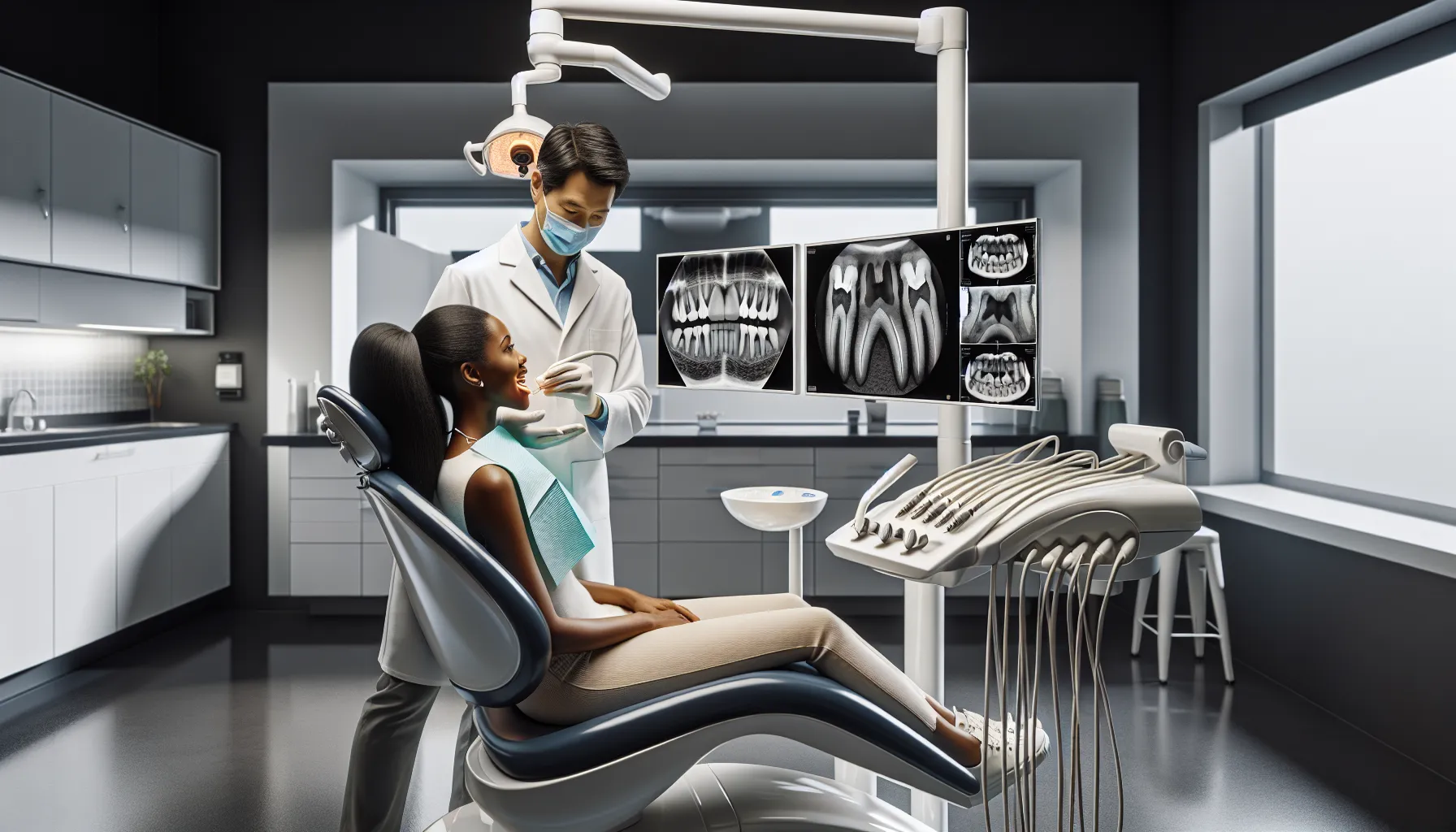
Think of dental X-rays as a window into the parts of your mouth we can’t see with our eyes alone. These diagnostic images capture detailed views of your teeth, bone, and the soft tissues around your jaw. When we do a routine exam, we’re checking surfaces, gums, and visible areas. But decay between teeth? Infections at the root? Bone density changes? That’s where X-rays come in.
The process itself is pretty straightforward. X-rays use low levels of ionizing radiation – sounds scary, but stick with us – to pass through your oral tissues. The film or digital sensor placed in or near your mouth registers these rays as they travel through different structures. Dense areas like teeth and bone absorb more radiation and show up lighter on the image, while softer tissues appear darker. The result? A detailed picture that shows us exactly what’s happening beneath the gum line.
What surprises most people is just how much information we get from a single image. We can spot early-stage cavities that haven’t caused symptoms yet, identify cysts or tumors, check the positioning of incoming teeth, and assess bone health around existing teeth. Without this technology, we’d basically be flying blind, waiting for problems to become obvious – and by then, treatment gets a lot more complicated.
Types of Dental X-Rays and Radiographic Imaging
Not all dental X-rays are created equal. We use different types depending on what we’re investigating, and understanding the options helps explain why we might recommend one over another. The main categories break down into intraoral (sensor or film inside your mouth) and extraoral (imaging from outside). Each serves a specific purpose.
Intraoral X-Rays
These are the ones you’re probably most familiar with – the small sensors or film pieces we position inside your mouth. They give us incredibly detailed views of individual teeth and the surrounding bone.
Bitewing X-rays are our go-to for detecting cavities and monitoring bone density. You bite down on a small wing-shaped sensor, and we capture images of your upper and lower back teeth in one shot. They’re perfect for spotting decay between teeth (where your toothbrush can’t reach) and checking the fit of crowns or fillings. If you’ve ever wondered why we ask about cavities you didn’t know you had, bitewings are usually how we found them.
Periapical X-rays zero in on one or two teeth at a time, showing everything from the crown down to the root tip and the surrounding bone. These are essential when we suspect a root infection, abscess, or bone loss around a specific tooth. If you’re experiencing pain in one area, this is often the first image we’ll take to figure out what’s going on.
Occlusal X-rays show the full arch of your upper or lower teeth. We use these less frequently, but they’re invaluable for locating extra teeth that haven’t erupted, identifying cysts, or assessing jaw fractures after an injury. Kids often get these as we track their dental development.
Extraoral X-Rays
When we need a bigger picture – literally – we turn to extraoral imaging. These techniques capture your entire jaw, skull, or facial structures in one image.
Panoramic X-rays are the wide-angle shots of dental imaging. In a single image, we can see all your teeth, both jaws, the temporomandibular joints (TMJ), sinuses, and nasal area. They’re incredibly useful for spotting impacted wisdom teeth, planning orthodontic treatment, or looking for tumors and cysts. You’ve probably seen the machine that rotates around your head while you bite down on a plastic piece – that’s panoramic imaging.
Cephalometric X-rays focus on the side profile of your skull and show the relationship between your teeth and jaw. Orthodontists rely heavily on these for treatment planning, especially when considering braces or other alignment procedures. They help us understand your bite pattern and facial structure in ways that impact both function and aesthetics.
Why Dental X-Rays Are Essential for Oral Health
We’ll be honest: if we could reliably diagnose and treat dental problems without X-rays, we would. But the reality is that some of the most serious oral health issues develop completely out of sight. By the time they’re visible or causing pain, we’re often looking at more invasive and costly treatment. That’s where radiographic imaging becomes essential.
Early Detection of Dental Problems
This is the big one. X-rays reveal problems before they become problems, if that makes sense. Cavities forming between teeth, infections brewing at the root, bone loss from gum disease – these conditions don’t announce themselves early on. You might feel perfectly fine while decay is quietly spreading or an abscess is forming.
We’ve seen it countless times: a patient comes in for a routine cleaning with zero complaints, and a bitewing X-ray shows a cavity that’s already halfway to the nerve. Catching it at that stage means a simple filling instead of a root canal later. Same with bone loss from periodontal disease. The earlier we spot it on X-rays, the better chance we have of stopping or slowing the progression.
X-rays also help us find issues that might never be visible during an exam – like cysts, tumors, or extra teeth that haven’t erupted. These conditions can cause serious complications if left undiagnosed, but they often show no symptoms in their early stages.
Treatment Planning and Monitoring
Even when we know what needs to be done, X-rays guide how we do it. Planning for dental implants? We need detailed images of your bone structure to determine placement and whether bone grafting is necessary. Considering braces or aligners? Orthodontists use X-rays to map out tooth positioning and predict how teeth will move.
Extractions, especially for impacted wisdom teeth, require X-rays to understand the tooth’s position relative to nerves and sinuses. Root canals need images to show us the number and shape of root canals in a tooth – it varies more than you’d think. Without that roadmap, treatment becomes guesswork.
We also use X-rays to monitor treatment over time. After a root canal, we’ll take follow-up images to make sure the infection has cleared and the bone is healing. If you’re at high risk for gum disease, periodic X-rays help us track bone levels and adjust your treatment plan as needed. It’s like having checkpoints along the way to make sure we’re heading in the right direction.
Safety and Radiation Exposure Concerns
Let’s address the elephant in the room: radiation. It’s completely reasonable to wonder whether dental X-rays are safe, especially if you or your kids need them regularly. The short answer? Yes, they’re safe. But let’s talk specifics because understanding the actual risk puts things in perspective.
Modern dental X-ray systems emit incredibly low levels of radiation. We’re talking minimal exposure, especially with digital sensors, which have further reduced radiation compared to traditional film. To put it in context, the radiation from a set of bitewing X-rays is less than what you’d naturally absorb from background sources during a typical day or two. A cross-country flight exposes you to more radiation than a full-mouth X-ray series.
That said, we don’t take radiation exposure lightly. We follow the ALARA principle – As Low As Reasonably Achievable. That means using lead aprons and thyroid collars when appropriate, positioning equipment correctly, and only taking images when there’s a clear diagnostic need. We’re not snapping X-rays for fun. Every image serves a purpose.
Digital radiography has been a game-changer here. Not only do digital sensors require up to 80% less radiation than traditional film, but the images appear instantly, so there’s no need for retakes due to processing errors. We can also enhance and manipulate digital images for better diagnosis without additional exposure.
If you’re pregnant, we’re extra cautious. While dental X-rays are generally considered safe during pregnancy – especially with proper shielding – we’ll often postpone non-urgent imaging until after delivery unless there’s an acute problem that needs immediate attention.
How Often Should You Get Dental X-Rays?
This is one of those “it depends” situations, which we know can be frustrating. But the truth is, there’s no one-size-fits-all schedule. How often you need X-rays depends on your age, current oral health, risk factors, and dental history.
Healthy adults with no history of cavities or gum disease typically need bitewing X-rays every one to two years. If your check-ups consistently show no problems and you practice good oral hygiene, we can space them out. It’s really about maintaining a baseline and catching any changes early.
Children and teenagers are a different story. Their mouths are constantly changing as teeth develop and erupt, so we often recommend more frequent imaging to monitor growth patterns and catch problems early. Kids are also more prone to cavities, so bitewings might be necessary every six months to a year depending on their decay risk.
If you’re dealing with active dental disease – whether that’s cavities, gum disease, or other ongoing issues – we’ll need X-rays more frequently to monitor treatment progress and catch new problems quickly. High-risk patients benefit from closer monitoring because early intervention is key to preventing more serious complications.
New patients typically get a full set of X-rays during their first visit so we can establish a complete baseline. This gives us a comprehensive view of your oral health and helps us compare changes over time. If you’re switching dentists and have recent X-rays from your previous office, we can often request those images to avoid unnecessary duplication.
Bottom line: we’re not going to recommend X-rays unless there’s a good clinical reason. Trust that we’re balancing the diagnostic benefits against any potential risks and tailoring the schedule to your individual needs.
Digital vs. Traditional Dental Radiography
If you haven’t been to the dentist in a while, you might be surprised by how much the technology has changed. Digital X-rays have largely replaced traditional film in most practices, and for good reason. The differences go beyond just convenience.
Digital X-rays use electronic sensors instead of film. The biggest advantage? Lower radiation exposure. Digital sensors are more sensitive than film, so they require significantly less radiation to capture a clear image. We’re talking about a 60-80% reduction in exposure compared to traditional film X-rays.
The images also appear instantly on our computer screens. No waiting for film to develop in a darkroom, no chemical processing, no chance of under- or over-exposure requiring a retake. We can see the image within seconds and immediately discuss findings with you. If we need a better angle or additional views, we know right away instead of discovering it after you’ve left.
Digital images are also incredibly versatile. We can zoom in on specific areas, adjust contrast and brightness for better visualization, and even use software to highlight or measure structures. Sharing images with specialists or transferring records to a new dentist is as simple as sending an email attachment. No more waiting for film copies to be mailed or physically transported.
Traditional film X-rays aren’t bad – they’ve served dentistry well for decades. But they do require higher radiation doses, take longer to process, and involve chemical waste from development. Storage is also more cumbersome since physical films take up space and can deteriorate over time.
Some practices still use film in certain situations, and that’s okay. But if you have a choice, digital offers clear advantages in terms of safety, efficiency, and diagnostic capability. It’s one of those technological upgrades that benefits everyone involved.
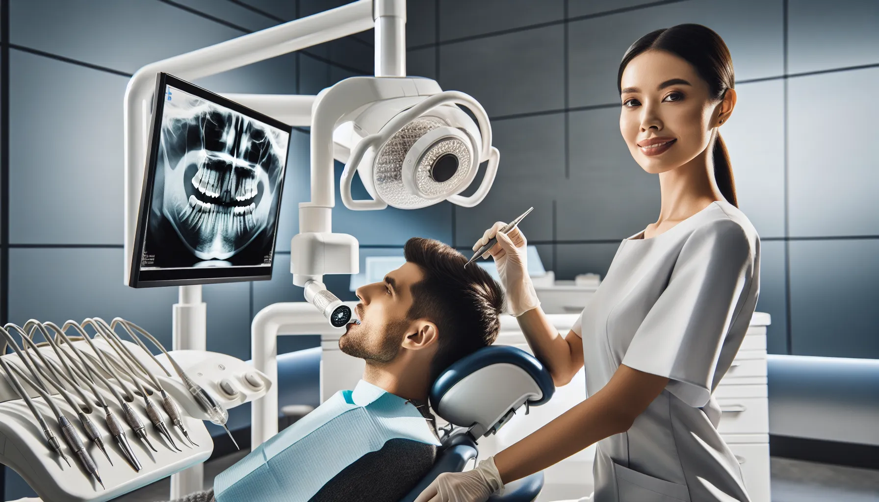
What to Expect During a Dental X-Ray Procedure
If you’ve never had dental X-rays before, or if it’s been a while, knowing what to expect can ease any anxiety. The process is quick, painless, and honestly pretty straightforward. We want you to feel comfortable, so here’s the typical walk-through.
First, we’ll have you settle into the dental chair. Depending on the type of X-ray, you might stay seated or stand (panoramic X-rays typically require standing with your chin on a rest). We’ll place a lead apron over your chest and lap to shield your body from any scatter radiation. Some setups include a thyroid collar for extra protection around your neck.
For intraoral X-rays, we’ll position a small sensor or film holder inside your mouth. We’ll ask you to bite down gently or hold it in place with your finger. It might feel a bit awkward, but it shouldn’t hurt. If you have a strong gag reflex, let us know. We can adjust the positioning or use techniques to make it more comfortable. Sometimes taking slow, deep breaths through your nose helps.
Once the sensor is positioned, we’ll step out of the room or behind a protective barrier. This isn’t because X-rays are dangerous to you – remember, the exposure is minimal – but because we’re taking images all day long, and cumulative occupational exposure adds up. We’ll ask you to stay very still for just a few seconds while the image is captured. You won’t feel or hear anything. Then we’ll reposition for the next image if needed.
Panoramic and cephalometric X-rays involve standing or sitting while a machine rotates around your head. You’ll bite down on a plastic piece to keep your head steady, and the machine does the rest. The rotation takes maybe 10-15 seconds, and again, you won’t feel anything.
The whole process, even for multiple images, usually takes less than 10 minutes. Digital X-rays mean we can review the images with you right away, pointing out any areas of concern or reassuring you that everything looks good. We’ll explain what we’re seeing and answer any questions before moving on to the rest of your appointment.
If you’re anxious about radiation, uncomfortable with things in your mouth, or have any other concerns, speak up. We’ve worked with all kinds of situations and can make adjustments to help you through it. The goal is getting the diagnostic information we need while keeping you as comfortable as possible.
Conclusion
Dental X-rays might not be the most exciting part of your dental visit, but they’re absolutely one of the most valuable. They give us the insight we need to catch problems early, plan effective treatments, and monitor your oral health over time. Without them, we’d miss hidden cavities, brewing infections, and bone loss until symptoms forced your hand – and by then, treatment is almost always more involved.
The good news? Modern radiographic imaging is safer, faster, and more effective than ever before. Digital technology has slashed radiation exposure while giving us clearer, more detailed images that we can analyze instantly. We’re not taking X-rays just to check a box. Every image serves a purpose, and we’re committed to using them responsibly.
So next time we mention taking X-rays, you’ll know exactly why. We’re not just looking at what’s visible on the surface. We’re making sure everything beneath it is healthy too. That’s how we keep your smile strong for the long haul.